Retinal Surgery
چهار شنبه 8 فروردین 1397
بازدید: 2638
Retinal surgeries

How are retinal tears treated?
Not all retinal breaks need to be treated. Many people have small holes in their retina that don't affect their vision and almost never produce associated symptoms. In general, however, if a retinal break is discovered in association with new symptoms or there is evidence of other high risk factors for a retinal detachment (previously described), one of two methods of treatment is indicated:
Laser Photocoagulation

Treatment spots are made around the tear to create a seal and keep fluid from accumulating underneath the retina.
Cryopexy
This technique freezes the area around the tear in order to seal the retinal break. This method is used when there is a large amount of blood and the laser cannot effectively reach the retina.
If retinal breaks are promptly identified, both treatments are highly successful in avoiding subsequent retinal detachment. Both of these procedures can be performed in all of The Retina Group of Washington offices.
It is unfortunate that some retinal tears progress to detachment almost immediately, sometimes without any symptoms. If this occurs, your retina specialist will recommend surgical repair.
How are retinal detachments treated?
Retinal detachments are most often repaired surgically by a retina specialist. Depending on the nature of your detachment, he or she will determine which specific procedure to pursue. Based on the variables, either one or a combination of the following operations is recommended:
Scleral Buckle Surgery

Scleral buckle surgery is a long established technique used to repair a retinal detachment. It is a method of closing breaks and flattening the retina. A small, invisible synthetic band, usually made of silicone rubber, is attached to the outside of the eyeball to gently push the wall of the eye against the detached retina. This relieves the traction caused by retinal breaks and also displaces some retinal fluid away from the break. Different sizes and types of scleral buckles may be used depending on the nature of the detachment.
Single operation success rates for scleral buckle, in general, are high. The operation is often performed as outpatient surgery and does not typically require an overnight hospital stay.
Following the procedure, your retina specialist may recommend you remain in a particular position during recovery in order to increase the chance of a successful result. You may experience some pain for a few days after the surgery; your eye may be swollen, red and/or tender for several weeks. You may be asked to put drops in your eye to help prevent infection and to keep the pupil from constricting. It may also be necessary to wear a patch over the eye at night.
A common side effect of scleral buckle surgery is increased nearsightedness. There are many less common complications that include eye infection, increased eye pressure, bleeding, injury to other parts of the eye, cataract, droopy eyelid and double vision. Complete vision loss is very rare, but possible as a result of scleral buckle surgery. As this procedure can change the shape of the eye, your prescription will likely change and you will ultimately need to have your contact lens or glasses prescription checked. If the initial operation is unsuccessful, additional surgery is likely to be recommended.
Pars Plana Vitrectomy
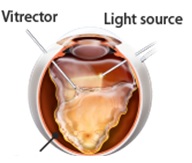
During a vitrectomy, the doctor makes three small incisions in the sclera (white of the eye). The vitreous, a gel-like substance that fills the eye, is removed using a small instrument. The fluid under the retina is then drained and laser treatment is applied to the tear and any other weak areas of the retina. Gas or silicone oil is then injected into the eye to replace the vitreous, reattach the retina and keep fluid from getting through the retinal break and detaching the retina again. Your doctor may ask you to position yourself face down or on your side in order to allow the gas or silicone oil to work most effectively to prevent fluid from getting under the retina during the early post-operative period.
Gas
During the healing process, the eye makes fluid that gradually replaces the gas. Because the gas is reabsorbed spontaneously, surgery to remove the gas is not needed. Different types of gas bubbles are used which can last in your eye for up to two months. Certain travel restrictions may apply while gas is present.
Silicone Oil
This may be used in the place of gas for more complex or recurrent detachments. Silicone oil does not reabsorb spontaneously and needs to be removed by a second surgery.
Regardless of whether gas or silicone oil is used, the single-operation success rate for vitrectomy is high. It is often performed on an outpatient basis and does not typically require an overnight hospital stay.
Complications are similar to those from scleral buckle surgery, even though the procedure doesn't always create more nearsightedness. It does, however, result more predictably in cataract formation. If this procedure is unsuccessful, your retinal specialist will likely recommend additional surgery.
Pneumatic Retinopexy
During a pneumatic retinopexy, a gas bubble is injected into the center of the eye to temporarily prevent fluid from entering through the retinal tear. Laser or cryopexy is then used to create a permanent seal. The specialized retinal pigment epithelial cells are then able to pump the existing fluid out from behind the retina. This procedure relies on the patient's ability to position their head so the small gas bubble stays over the retinal tear. For this reason, pneumatic retinopexy may not be the appropriate procedure for all retinal detachments. In select circumstances, the success rate of this procedure is high.
Pneumatic retinopexy is typically done in your retina specialist's office and can be performed at all of The Retina Group of Washington locations. As with other surgical procedures to repair retinal detachments, this has potential complications that include new post-operative retinal breaks, increased eye pressure, infection, gas getting behind the retina and cataract formation. Total visual loss is rare, but possible following this operation. Here again if this procedure is unsuccessful, your doctor will likely recommend additional surgery.
Medical Health
Depending on your overall health, your retina specialist may ask you to see your primary care provider for a general health check prior to retina surgery. Be sure to let your surgeon know if you have any serious medical conditions or allergies and if you are on blood thinners or other medications that slow clotting of your blood.
دیدگاه های ارسال شده توسط شما، پس از تایید مدیر سایت در وب سایت منتشر خواهد شد.
پیام هایی که حاوی تهمت یا افترا باشد منتشر نخواهد شد.
پیام هایی که به غیر از زبان فارسی یا غیر مرتبط با خبر باشد منتشر نخواهد شد.