Paraclinical Servises
چهار شنبه 8 فروردین 1397
بازدید: 2753
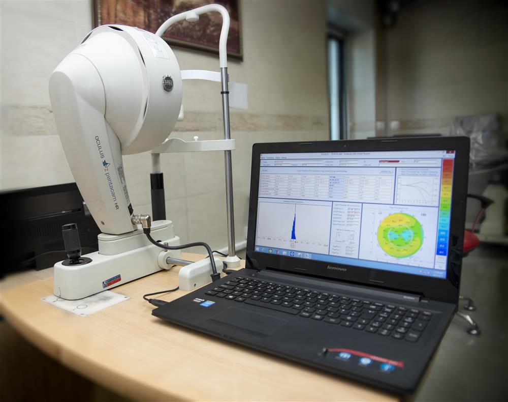
Pentacam
Combined device consisting of a slit illumination system and a Scheimpflug camera which rotates around the eye to give an automatic measurement of an over-all view of the anterior segment of the eye. It provides topographic data on elevation and curvature of the entire anterior and posterior corneal surface, used also for kerataconus detection and monitoring and glaucoma screening.
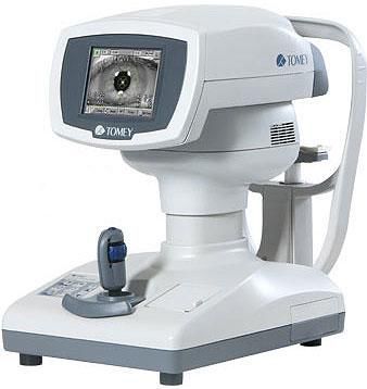
Specular Endothelial Camera
A camera which measures the density of the corneal endothelial cells which is necessary before corneal transplantation and other corneal procedures.
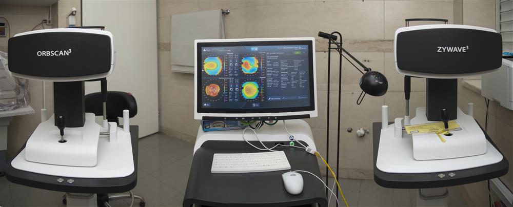
Orbscan
The Orbscan is an advanced diagnostic device that quickly and painlessly measures the thickness and shape of both the front and back surfaces of the cornea, providing a three-dimensional map of the complete corneal structure. The test is an essential first step to check for irregularities in your eyes or other conditions that may limit candidacy or compromise the safety of the LASIK procedure. If the Orbscan® measurements show that you are a good cand idate for LASIK, your surgeon can create a customized surgical plan for you.

Corneal Topography
It is a noncontact examination that photographs the surface of the eye using ordinary light. The greatest advantage of corneal topography is its ability to detect conditions invisible to most conventional testing.

Aberrometry
Corneal aberrometry is a test that measures the way light travels through the entire optical pathway, and compares it to the way light travels through an optically perfect eye.
This assessment is often done using a machine called a wave front aberrometer. By bouncing light off the retina and measuring the imperfections of the eye, the wave front aberrometer creates a three dimensional map of the cornea.
This corneal map is then used for pre-operative screening to design a customized ablation, or tissue removal, that addresses the patient’s unique aberrations, or imperfections. Customized ablation is used in both PRK and LASIK procedures. On surgery day, the information from the map is transferred electronically to the laser, enabling the provider to customize a laser vision procedure to the patient’s particular needs.
Pachymetry - A Simple Test to Determine Corneal Thickness
A pachymetry test is a simple, quick, painless test to measure the thickness of your cornea. With this measurement, your doctor can better understand your IOP reading, and develop a treatment plan that is right for your condition. The procedure takes only about a minute to measure both eyes.

Ocular Response Analyzer® (ORA) is the only tonometer that measures Corneal Hysteresis (CH), a superior predictor of glaucoma progression. Corneal Hysteresis is an indication of the biomechanical properties of the cornea differing from thickness or topography, which are geometrical attributes. in addition to Corneal Hysteresis, Ocular Response Analyzer provides Corneal Compensated Intraocular Pressure (IOPcc), a better indication of the true pressure, proven to be less influenced by corneal properties than Goldmann or other methods of tonometry.2

Posterior OCT- MRW (Nerve Fiber Layer Analysis)
A non-contact measurement of the thickness of the nerve fibre layer. It is used in the early detection and the assessment of progression of glaucoma.
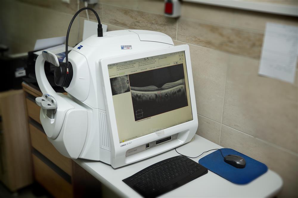
Posterior OCT-Heidelberg
A non-contact precise measurement of the retina (posterior pole), retinal pigment epithelium and choroid. It is used in the management macular disease (especially macular holes and macular degeneration), diabetic eye disease and the assessment of reduced vision.

Anterior OCT
A non-contact precise measurement of the anterior segment of the eye including cornea, drainage angle, anterior chamber, iris and anterior lens surface. It is used in the management of glaucoma, uveitis, difficult cataracts, complicated cataract surgery and other diseases of the anterior chamber.

Fluorescein Angiography-Heidelberg
Digital imaging of the retina (and occasionally sclera and iris) following the intravenous injection of fluorescein to assess perfusion and vascular integrity. It is used particularly in the management of age related macular degeneration and diabetes.

Visual Field Test
An automated test of peripheral visual field in a reproducible and internationally recognised format.
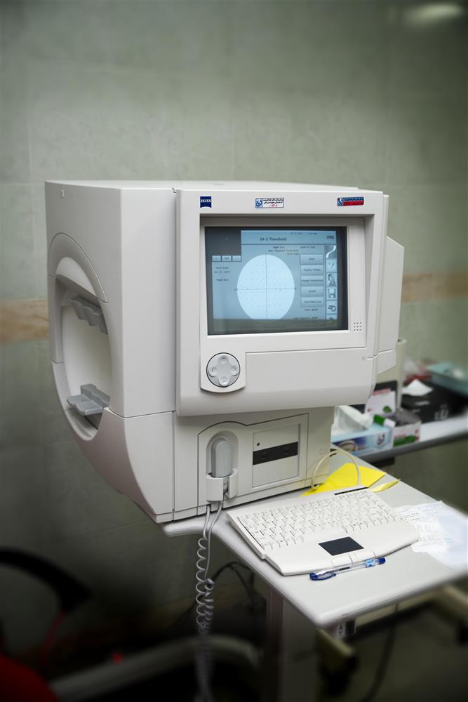
Specular Endothelial Camera
A camera which measures the density of the corneal endothelial cells which is necessary before corneal transplantation and other corneal procedures.
IOL Power measurement (IOL master)
IOL master
A non-contact measurement of the length of the eye and size / shape of the cornea to calculate the strength of the lens implant needed to correct vision after cataract extraction.
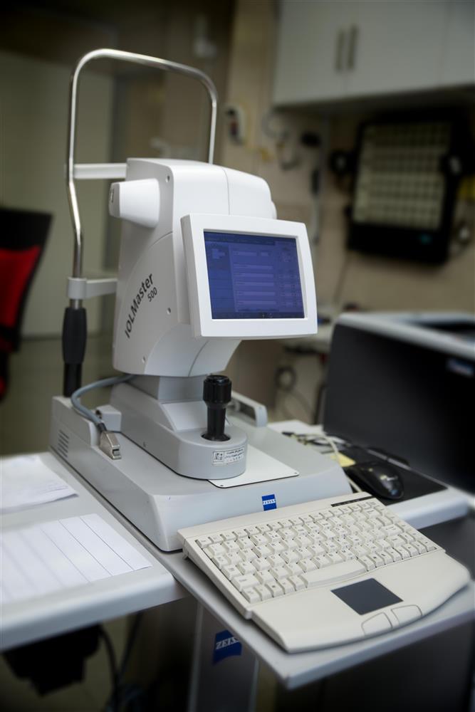
Cataract biometry
A contact measurement of the length of the eye and size / shape of the cornea to calculate the strength of the lens implant needed to correct vision after cataract extraction.
Ultrasonography
Imaging of the inside of the eye using ultrasound, it is used where the view inside the eye is obscured for whatever reason, or to measure the size of features inside the eye. It is often used in the assessment of diabetic eyes before surgery.

Contrast sensitivity
contrast sensitivity is a very important measure of visual function, especially in situations of low light, fog or glare, when the contrast between objects and their background often is reduced. Driving at night is an example of an activity that requires good contrast sensitivity for safety.

دیدگاه های ارسال شده توسط شما، پس از تایید مدیر سایت در وب سایت منتشر خواهد شد.
پیام هایی که حاوی تهمت یا افترا باشد منتشر نخواهد شد.
پیام هایی که به غیر از زبان فارسی یا غیر مرتبط با خبر باشد منتشر نخواهد شد.